Scatter and Collimation
 |
 |
 |
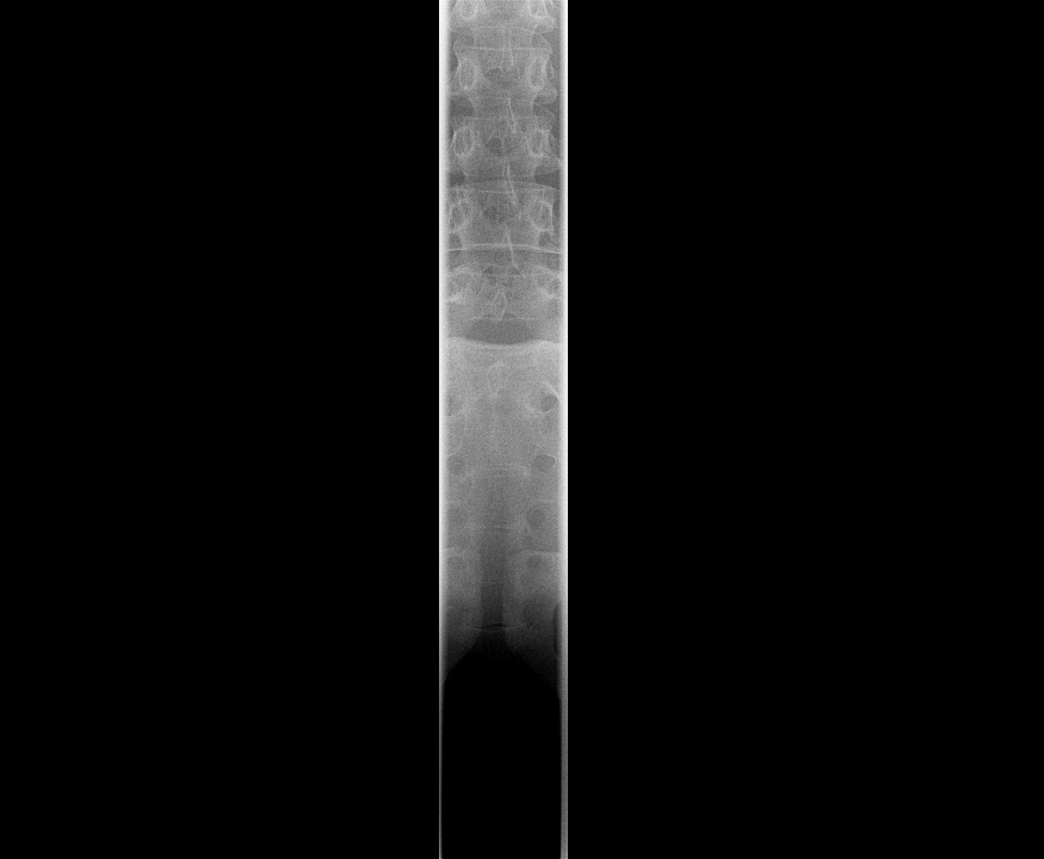 |
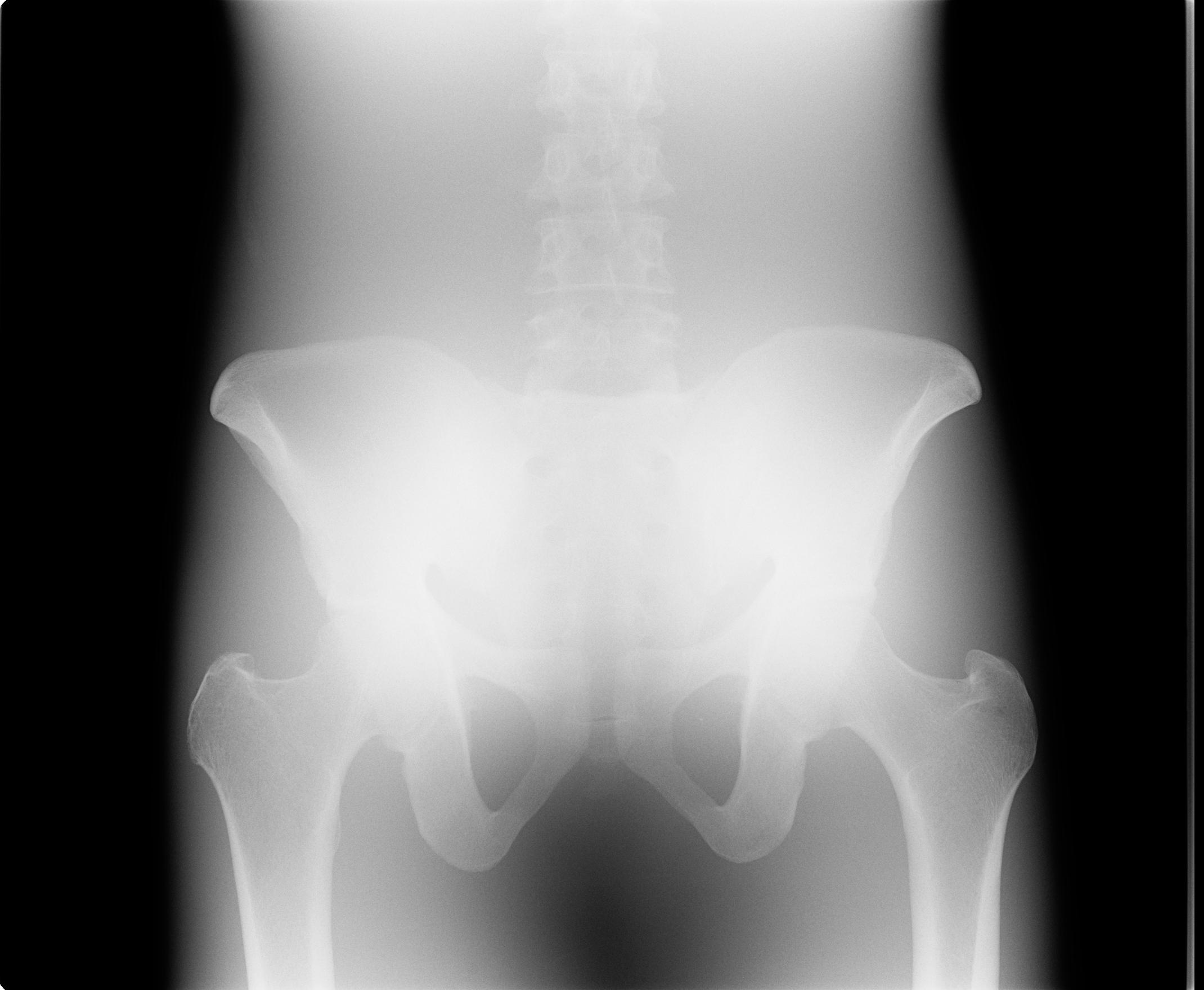
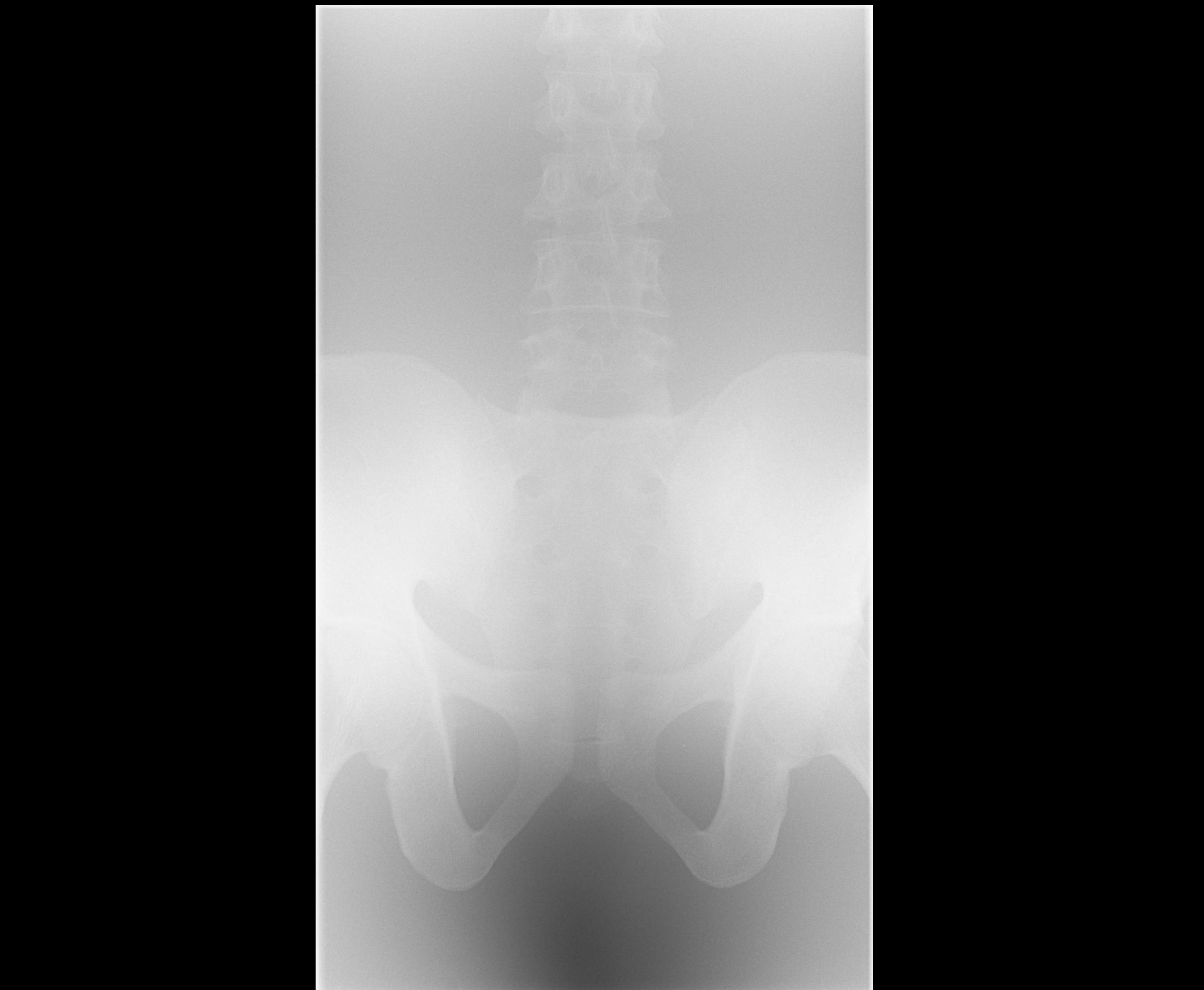
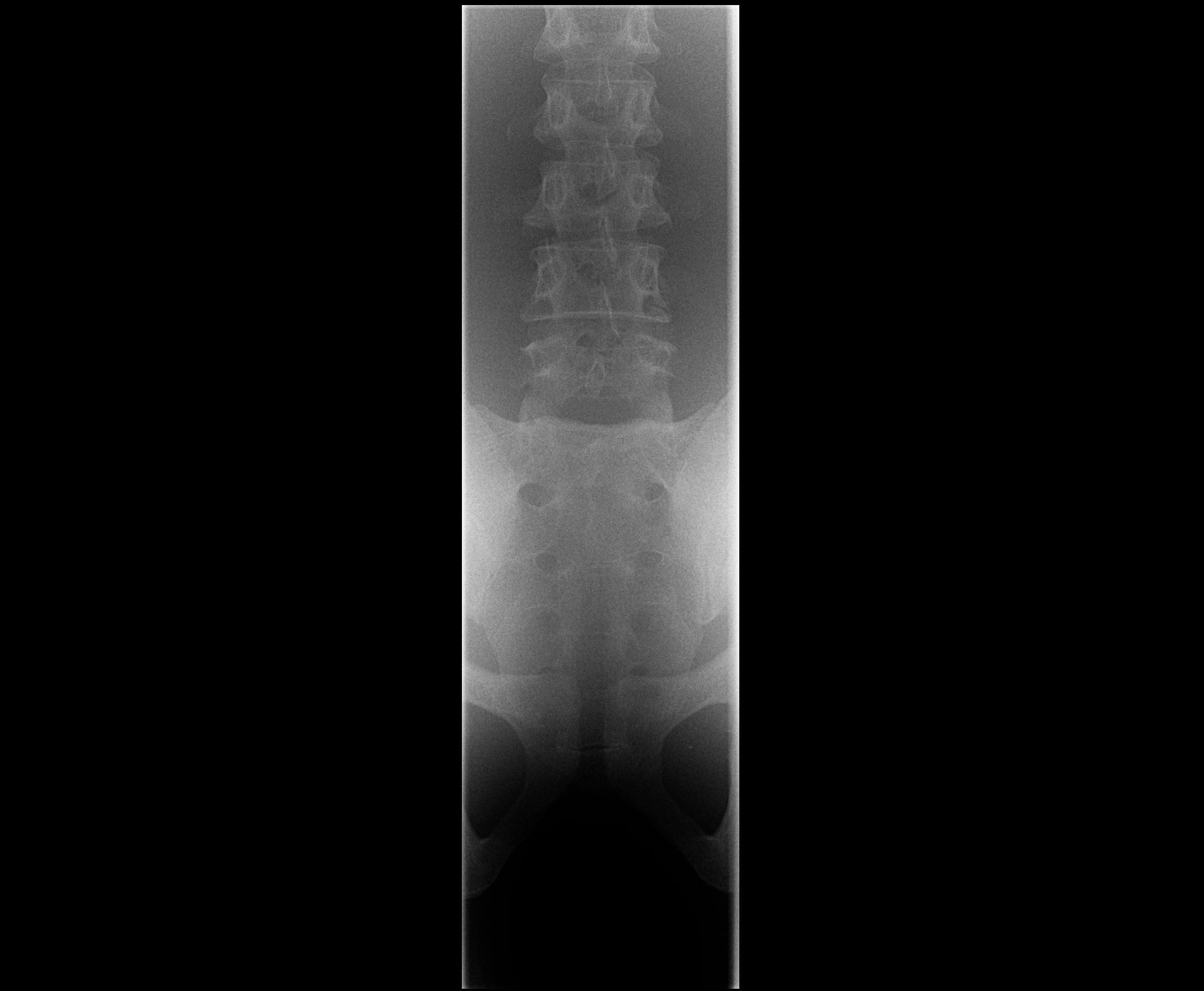
The set of images of the pelvis phantom shown above were all obtained using the same radiographic technique (70 kV and 3 mAs), and obtained using a computed radiography cassette with no scatter removal grid. The four successive images of the pelvis phantom, used lateral dimensions of 43 cm (upper left), 20 cm (upper right), 10 cm (lower left), and 5 cm (lower right). As the collimation is narrowed, the visibility of the spine markedly improves because scatter generated in the phantom has an increasingly likely chance of missing the exposed image region. This example illustrates that any narrow irradiation geometry, such as that used in CT, will be effective in removing scatter. It also shows that scatter is very important issue when large areas are exposed, and for conventional radiography, it is essential to take steps to minimize the amount of scatter reaching the image receptor.